

Equipment
We charge time-dependent costs for the use of BCF devices. The costs (net prices per hour) are shown in a table (Costs). No other costs for maintenance, repairs or replacement are charged.
- Zeiss Spinning Disc Cell Observer, inverted
- Zeiss LSM780-Airyscan, inverted
- Leica TCS SP8, inverted
- Leica TCS SP8 Falcon, inverted
- Leica TCS SP5 STED, inverted
- Zeiss LSM 700, inverted
- LSM 800, upright
- Zeiss Imager.Z2 ApoTome.2, upright
- Axioplan 2 Imaging
- IncuCyte Zoom
- AxioObserver.Z1, inverted
- Zeiss Axiophot 2e, upright
- Calcium Imaging
- Zeiss Axioscan 7
- Zeiss PALM Microbeam - Laser Microdissection, inverted
- AFM JPK Nanowizard 3, inverted
- Lenovo Graphics Workstation
- HP Z4 Graphics Workstation
- HP Z6 Graphics Workstation
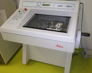
- Cryostat, Leica 3050S
- Microtom Leica RM2255
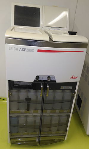
- Embedding, Leica ASP 200 S
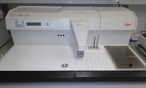
- Embedding, Leica EG1160
2.) Instruments not managed by the BCF
tba
Spinning Disc Microscope (Confocal)
Zeiss Spinning Disc Cell Observer, inverted, GenTSV-S1 Lab, AG Cell Biology, SIKT Building
| Instrument: | Zeiss Cell Observer SD System, Spinning Disc- Yokohama CSU-X1 , inverted, FRAP module |

|
||||||||||||||||||||||||||||
|---|---|---|---|---|---|---|---|---|---|---|---|---|---|---|---|---|---|---|---|---|---|---|---|---|---|---|---|---|---|---|
| Illumination: |
|
|||||||||||||||||||||||||||||
| Scan: | Spinning Disc, disc speed 1.500-5.000 U/min | |||||||||||||||||||||||||||||
| Objectives: |
|
|||||||||||||||||||||||||||||
| Incubator: | Incubator XL multi S1 Dark LS for Live Cell Imaging | |||||||||||||||||||||||||||||
| Filter: |
|
|||||||||||||||||||||||||||||
| Others: | Confocal in vivo microscopy, FRAP, Z-stacks, time-series, mosaic scan | |||||||||||||||||||||||||||||
Laser Scanning Microscopes (Confocal)
Zeiss LSM780-Airyscan, inverted, GenTSV-S1 Lab, AG Cell Biology, SIKT Building
| Instrument: | LSM 780 inverted, Airyscan-Module (Super Resolution Microscopy) 34 channel Quasar Detector, FRAP module, mosaic scan and time series etc. |
 34 channel Quasar Detector, FRAP module, mosaic scan and time series etc.")
|
|---|---|---|
| Stative: | Axio Observer.Z1 inverted motorized (steps 10 nm), TFT-Touchscreen | |
| Illumination: |
|
|
| Airyscan: | 1,7 x improved resolution | |
| Detector: | GaAsP | |
| Objectives: |
|
|
| Others: | Super Resolution Microscopy (verified 110nm XY scan resolution by Zeiss service), Z-stacks, time-series, mosaic scan, FRAP |
Leica TCS SP8, inverted, GenTSV-S2 Lab, VMF Campus / Vet.-Anat.-Inst.
| Instrument: | Leica TCS SP8 DMi8, inverted |

|
|---|---|---|
| Illumination: | 405, 458, 476, 488, 496, 514, 561, 633 nm | |
| Scan mode: | 10, 100, 200, 400, 600, 700, 1000, 1400, 1800, 8000 Hz | |
| Beamsplitter: | Acusto-optic beam splitter (AOBS), detection range 400 bis 800 nm in 1 nm steps | |
| Detector: |
|
|
| Objectives: |
|
|
| Others: |
|
Leica TCS SP8 Falcon, inverted, GenTSV-S1 Lab, AG BioPhysicalChemistry, Johannisallee 21
| Instrument: | Leica TCS SP8 FALCON |

|
|---|---|---|
| Illumination: |
|
|
| Scan mode: | 10, 100, 200, 400, 600, 700, 1000, 1400, 1800, 8000 Hz | |
| Beamsplitter: | Acusto-optic beam splitter (AOBS) detection range 400 to 800 nm in 1 nm steps | |
| Detector: |
|
|
| Objectives: |
|
|
| Others: |
|
Leica TCS SP5 STED, inverted, Talstr. 33
| Instrument: | Leica TCS SP5 STED, DMi6 inverted |

|
|---|---|---|
| Illumination: |
|
|
| Scan mode: | 10 - 1400 Hz | |
| Beamsplitter: | Acusto-optic beam splitter (AOBS) detection range 400 to 800 nm in 1 nm steps | |
| Detector: |
|
|
| Objectives: |
|
|
| Others: |
|
Zeiss LSM 700, inverted, GenTSV-S1 Lab, AG BioPhysicalChemistry, Johannisallee 21
| Instrument: | Zeiss LSM 700, AxioObserver.Z1, inverted, motorized |

|
|---|---|---|
| Illumination: | 405, 488, 555, 639 nm | |
| Beamsplitter: |
|
|
| Resolution: | maximal (2048 x 2048) Pxl | |
| Scan speed: | max. 5 frames/s @ (512x512) Pxl | |
| Detector: | 2x PMT for parallel detection of 2 channels | |
| Objectives: |
|
|
| Others: |
|
LSM 800, upright, Gen TSV-S1 Lab, AG Neurogenetics, Talstr. 33
| Instrument: | Zeiss LSM800 upright |

|
|---|---|---|
| Illumination: | Laser 405nm, 488nm, 561nm, 640nm | |
| Detector: | GaAsP | |
| Objectives: |
|
|
| Others: | Incubation system for Live Cell Imaging |
Widefield Structured Illumination
Zeiss Imager.Z2 ApoTome.2, upright, GenTSV-S1 Lab, AG Cell Biology, SIKT Building
| Instrument: | Zeiss Imager.Z2 ApoTome.2, Grid technology, deconvolution Axiocam 506 Color and AxioCamMrm, fully motorized stage, Definite Focus, Z-stacks, time series, mosaic scan |

|
||||||||||||||||||||||||||||||||||||||||||||||||||||
|---|---|---|---|---|---|---|---|---|---|---|---|---|---|---|---|---|---|---|---|---|---|---|---|---|---|---|---|---|---|---|---|---|---|---|---|---|---|---|---|---|---|---|---|---|---|---|---|---|---|---|---|---|---|---|
| Illumination: | Colibri: LED and HXP120 | |||||||||||||||||||||||||||||||||||||||||||||||||||||
| Objectives: |
|
|||||||||||||||||||||||||||||||||||||||||||||||||||||
| Filter: |
|
|||||||||||||||||||||||||||||||||||||||||||||||||||||
Widefield Epifluorescence Microscopy
IncuCyte Zoom, GenTSV-S2 Lab, AG Cell Biology, SIKT Building
| Instrument: | IncuCyte Zoom® Live Cell Imaging |
|---|---|
| Illumination: | LED |
| Objectives: |
|
| Filter: | GFP,RFP |
| Others: | In vivo microscopy, multi-well, incubator,IncuCyte Cell Migration Kit for Scratch and Wound- Assays |
AxioObserver.Z1, inverted, GenTSV-S1 Lab, AG BioPhysicalChemistry, Johannisallee 21
| Instrument: | Zeiss AxioObserver.Z1, Epifluorescence, inverted, motorized, transmission |

|
|---|---|---|
| Illumination: | Colibri LED; @ 365, 470, 555, 625 nm | |
| Camera: | Axiocam MRm3, (1388x1040) Pxl | |
| Objectives: |
|
|
| Filter: | DAPI, FITC, TRITC, Cy5, DIC (Transmission) | |
| Others: |
|
Zeiss Axiophot 2e, upright, GenTSV-S1 Lab, AG Cell Biology, SIKT Building
| Instrument: | Zeiss Axiophot 2e, Axioplan, Upright/ Fluorescence / bright-field / phase contrast |

|
|---|---|---|
| Illumination: | HXP 120C | |
| Objectives: |
|
|
| Filter: | bandpass/longpass filters to combine standard fluorescent dyes (DAPI, Cy2, Cy3, Cy5 etc.), see Apotom HXP filters | |
| Others: | imaging via Axiocam Hrc and Axiocam Hrm, standard fluorescent and bright-field microscopy including phase contrast microscopy |
Calcium Imaging, GenTSV-S1 Lab, AG Neurogenetics, Talstr. 33
| Instrument: | Zeiss AxioExaminer, upright |

|
|---|---|---|
| Illumination: | HXP and Colibri LED 385nm, 470nm, 555nm, 630nm | |
| Objectives: |
|
Axio Slide Scanner
Zeiss Axioscan 7, VMF Campus / Vet.-Patho.-Inst.
| Instrument: | Zeiss Axioscan 7 Slide Scanner for Brightfield |

|
|---|---|---|
| Camera: | Axioscan 7 KMAT | |
| Objectives: |
|
|
| Workstation: |
Workstation Premium ZEISS 60A R2 (hp Z6):
|
|
| Storage: |
|
|
| Link: | https://www.vetmed.uni-leipzig.de/institut-fuer-veterinaer-pathologie/institut/team |
Microdissection
Zeiss PALM Microbeam - Laser Microdissection, inverted, GenTSV-S1 Lab, AG Cell Biology, SIKT Building
| Instrument: | Zeiss Cell Observer Z.1, inverted, allows Laser based Microdissection |

|
||||||||||||||||||||||||||||
|---|---|---|---|---|---|---|---|---|---|---|---|---|---|---|---|---|---|---|---|---|---|---|---|---|---|---|---|---|---|---|
| Illumination: | Colibri.2 and HXP120 | |||||||||||||||||||||||||||||
| Objectives: |
|
|||||||||||||||||||||||||||||
| Filter: |
|
|||||||||||||||||||||||||||||
Atomic Force Microscopy
AFM JPK Nanowizard 3, inverted, GenTSV-S1 Lab, AG BioPhysicalChemistry, Johannisallee 21
| Instrument: | JPK Nanowizard 3, CellHesion-Modul, inverted: AxioObserver.D1 |

|
|---|---|---|
| Illumination: | Monochromator Polychrom IV (400-700nm) | |
| Camera: | Monochrom-Cam (Hamamatsu ORCA ERG , 1344x1024) | |
| Scan: | xy: 100x100µm , z: 15µm (CellHesion: z: 100µm) | |
| Untersuchungsmodi: |
Imaging:
|
|
| Noise Level: |
|
|
| Scan mode: | 0,1Hz – 1kHz | |
| Filter: | u.a. 470 / 515 LP, 546 / 590 LP | |
| Others: |
|
Image Analysis
Lenovo Graphics Workstation, VMF Campus
| Analysis-Software: |
|
|---|
HP Z4 Graphics Workstation, GenTSV-S1 Lab, SIKT Building
| Analysis-Software: |
|
|---|
HP Z6 Graphics Workstation, VMF Campus
| Analysis-Software: |
|
|---|
Histo: Cryostat / Microtom / Paraffin-Embedding
Cryostat, Leica 3050S, GenTSV-S1 Lab, AG Cell Biology, SIKT Building
| Link: | https://www.leicabiosystems.com/de/histologiegeraete/gefrierschnitte |
|
|---|
Microtom Leica RM2255, GenTSV-S1 Lab, AG Cell Biology, SIKT Building
| Link: | https://www.leicabiosystems.com/de/histologiegeraete/mikrotome/products/leica-rm2255/ |
|---|
Embedding, Leica ASP 200 S, GenTSV-S1 Lab; AG Cell Biology SIKT Building
| Link: | https://www.leicabiosystems.com/de/histologiegeraete/gewebeinfiltration/products/leica-asp200 |
|
|---|
Embedding, Leica EG1160, GenTSV-S1 Lab, AG Cell Biology, SIKT Building
| Link: | https://www.leicabiosystems.com/de/histologiegeraete/einbetten/products/leica-eg1160/ |
|
|---|